2 article(s) from Krzywiecki, Maciej
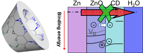
Cyclodextrin inhibits zinc corrosion by destabilizing point defect formation in the oxide layer
- Abdulrahman Altin,
- Maciej Krzywiecki,
- Adnan Sarfraz,
- Cigdem Toparli,
- Claudius Laska,
- Philipp Kerger,
- Aleksandar Zeradjanin,
- Karl J. J. Mayrhofer,
- Michael Rohwerder and
- Andreas Erbe
Beilstein J. Nanotechnol. 2018, 9, 936–944, doi:10.3762/bjnano.9.86


Impact of air exposure and annealing on the chemical and electronic properties of the surface of SnO2 nanolayers deposited by rheotaxial growth and vacuum oxidation
- Monika Kwoka and
- Maciej Krzywiecki
Beilstein J. Nanotechnol. 2017, 8, 514–521, doi:10.3762/bjnano.8.55


Other Beilstein-Institut Open Science Activities